AUTHOR
MATT JONES, MD
CONSULTANTS
KYLE DEAL, MD (INTERVENTIONAL RADIOLOGY)
JESSICA BURGESS, MD (TRAUMA SURGERY)
Don Byars, MD (EMERGENCY MEDICINE)
Barry Knapp, MD (EMERGENCY MEDICINE)
CONTRIBUTORS
MATT JONES, MD
DON BYARS, MD
ANNA KATE DEAL, MD
TAMARA ARMSTRONG, MD
CHARLES GRAFFEO, MD
DAVID NESBITT, MD
D. TAYLOR GAMMONS, MD
GIORGI TSEREDIANI, MD
LOGAN MCNEIVE, MD
CODY MCILVAIN, MD CANDIDATE CLASS OF 2020
JUSTIN YAWORSKY, MD CANDIDATE CLASS OF 2020
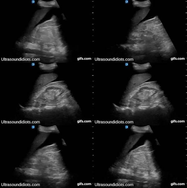
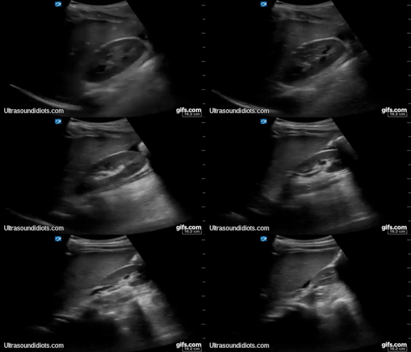
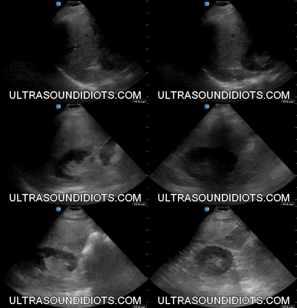
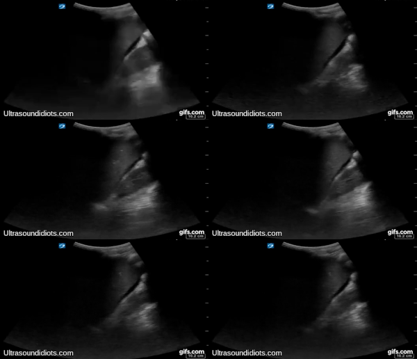
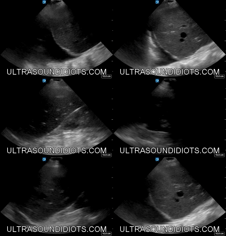
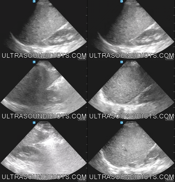
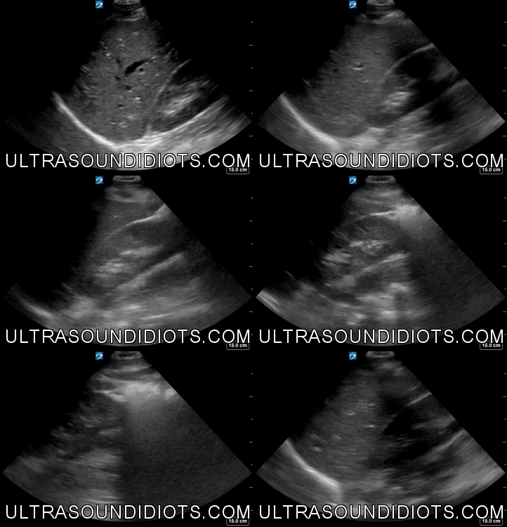
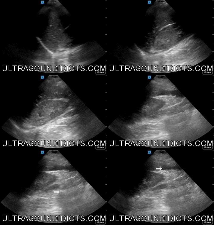
EFAST RUQ EXAM ACHIVE
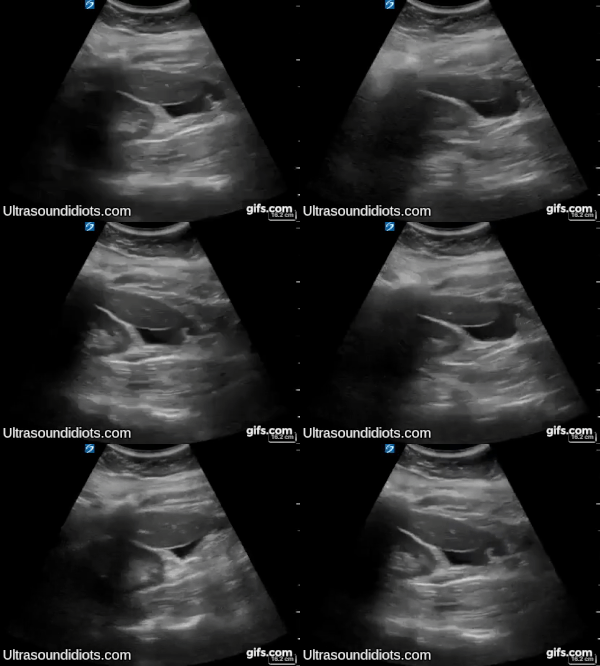
Exam 1
Probe: Curvilinear
Depth: Appropriate
Gain: Appropriate
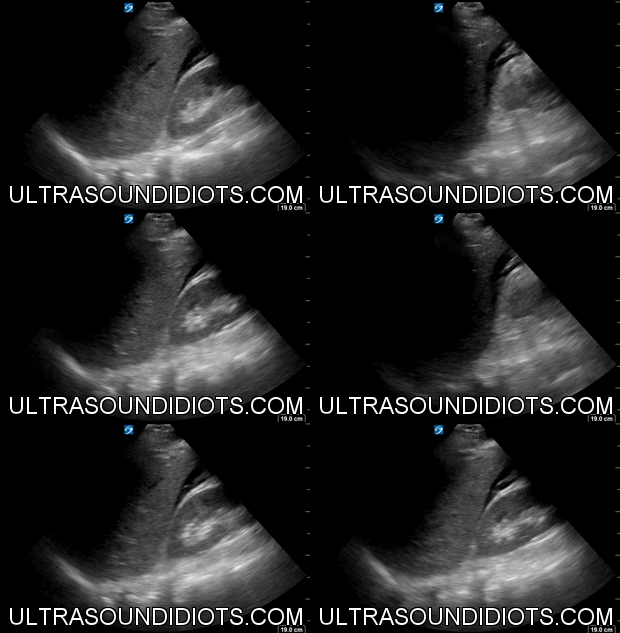
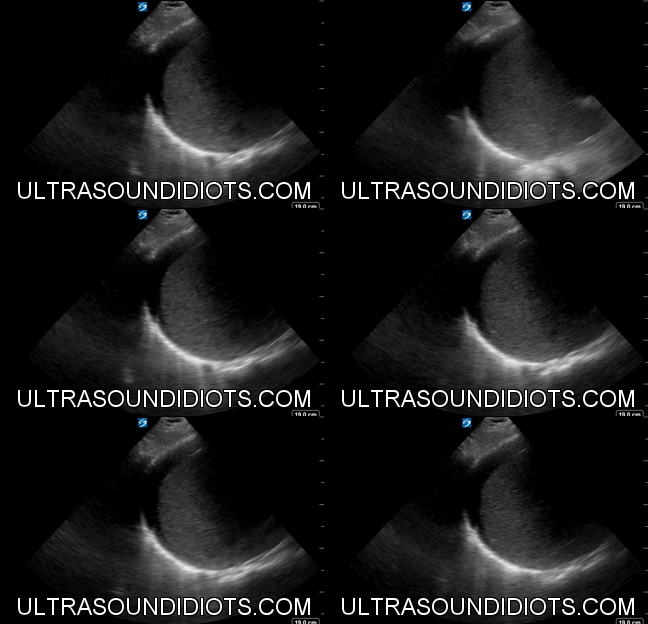
Findings: Free fluid located in the paracolic gutter (below the inferior pole of the right kidney). This location is often overlooked and can be easily missed if the sonographer does not slide the probe inferiorly. The pleural space, the subphrenic space (between diaphragm and liver), and the hepatorenal space (Morrison’s pouch) are not visualized in the clip.
Impression: Positive Exam, Incomplete Visualization of RUQ
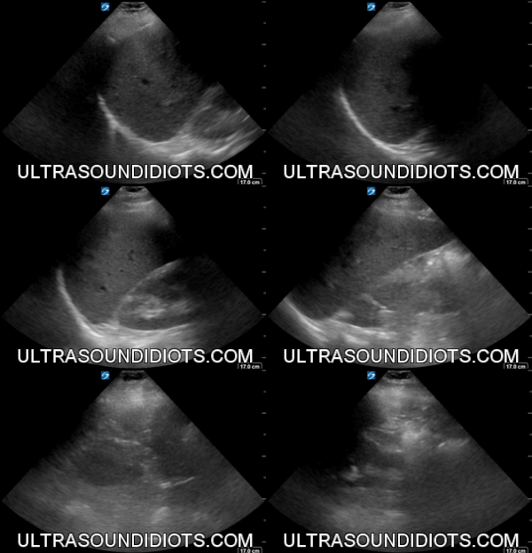
exam 2
Probe: Phased Array
Depth: Appropriate
Gain: Appropriate
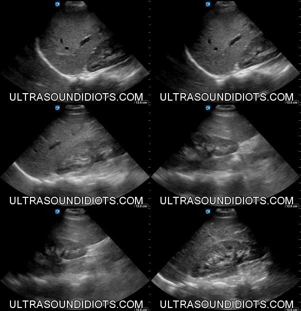
Findings: This is an excellent clip which clearly shows all four areas of interest- the pleural space, the subphrenic space (between diaphragm and liver), the hepatorenal space (Morrison’s pouch), and the paracolic gutter (inferior pole of the kidney).
Impression: Negative Exam, Complete Visualization of RUQ
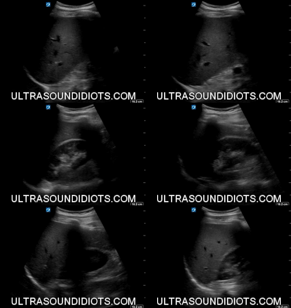
exam 3
Probe: Curvilinear
Depth: Appropriate
Gain: Too Low
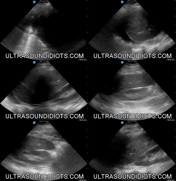
Findings: This clip does not adequately visualize the pleural space (space above the diaphragm). Otherwise, the exam shows no evidence of free fluid in the subphrenic space, the hepatorenal space or the inferior pole of the right kidney.
Impression: Indeterminate Study, Incomplete Visualization of RUQ
Exam 4
Probe: Curvilinear
Depth: Appropriate
Gain: Appropriate
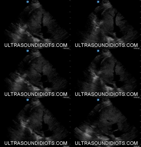
Findings: Free Fluid located within the hepatorenal space. The pleural space, subphrenic space and paracolic gutter are not visualized.
Impression: Positive Exam, Incomplete Visualization of RUQ
Exam 5
Probe: Curvilinear
Depth: Appropriate
Gain: Appropriate
Findings: Free Fluid located within hepatorenal space. The pleural space, subphrenic space and paracolic gutter are not visualized.
Impression: Positive Exam, Incomplete Visualization of RUQ
Exam 6
Probe: Phased Array
Depth: Appropriate
Gain: Appropriate
Findings: This clip does not adequately visualize the pleural space. Otherwise, the exam demonstrates no evidence of free fluid in the subphrenic space, hepatorenal space or paracolic gutter.
Impression: Indeterminate Exam, Incomplete Visualization of RUQ
Exam 7
Probe: Curvilinear
Depth: Appropriate
Gain: Too Low
Findings: The pleural and subphrenic spaces are not visualized. Free fluid is demonstrated within hepatorenal space. The paracolic gutter is not visualized.
Impression: Positive Exam, Incomplete Visualization of RUQ
EXAM 8
Probe: Phased Array
Depth: Appropriate
Gain: Appropriate
Findings: The pleural and subphrenic spaces are not visualized. Free fluid is demonstrated within hepatorenal space. The paracolic gutter is not visualized.
Impression: Positive Exam, Incomplete Visualization of RUQ
EXAM 9
Probe: Curvilinear
Depth: Appropriate
Gain: Too Low
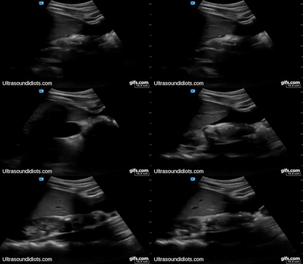
Findings: The pleural and subphrenic spaces are not visualized. The hepatorenal space is incompletely visualized. The gallbladder is present at the beginning of the clip and should not be confused with free fluid. Close inspection of the later part of the clip shows free fluid within the paracolic gutter.
Impression: Positive Exam, Incomplete Visualization of RUQ
EXAM 10
Probe: Curvilinear
Depth: Appropriate
Gain: Appropriate
Findings: There is fluid in the pleural space. The subphrenic space demonstrates no free fluid. The hepatorenal space and the paracolic gutter are not visualized
Impression: Positive Exam, Incomplete Visualization of RUQ
EXAM 11
Probe: Curvilinear
Depth: Appropriate
Gain: Appropriate
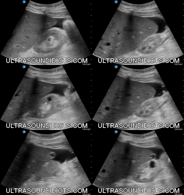
Findings: The pleural space and the subphrenic space are not visualized. There is no free fluid visualized with the hepatorenal space. There is free fluid demonstrated in the paracolic gutter. There is a benign (minimally complex, 1 small septum; Bosniak 2) cyst within the inferior pole of the right kidney.
Impression: Positive Exam, Incomplete Visualization of RUQ
EXAM 12
Probe: Phased Array
Depth: Appropriate
Gain: Appropriate
Findings: No free fluid in pleural space. Free fluid is demonstrated in the subphrenic space. The hepatorenal space and paracolic gutter are not visualized.
Impression: Positive Exam, Incomplete Visualization of RUQ
EXAM 13
Probe: Phased Array
Depth: Appropriate
Gain: Appropriate
Findings: The exam demonstrates no evidence of free fluid in the pleural space, subphrenic space, hepatorenal space or paracolic gutter.
Impression: Negative Exam, Complete Visualization of RUQ
EXAM 14
Probe: Phased Array
Depth: Appropriate
Gain: Appropriate
Findings: There is free fluid visualized within the pleural space. No free fluid is demonstrated within the subphrenic space, the hepatorenal space or the paracolic gutter..
Impression: Positive Exam, Complete Visualization of RUQ
EXAM 15
Probe: Phased Array
Depth: Appropriate
Gain: Too Low
Findings: There is fluid in the pleural space. The subphrenic space demonstrates no free fluid. The hepatorenal space and the paracolic gutter are not visualized
Impression: Positive Exam, Incomplete Visualization of RUQ
EXAM 16
Probe: Curvilinear
Depth: Appropriate
Gain: Appropriate
Findings: The pleural space is inadequately visualized. There is no fluid within the subphrenic space. The right kidney is absent (s/p right nephrectomy). No fluid is demonstrated within the hepatorenal space or the paracolic gutter.
Impression: Negative Exam, Complete Visualization of RUQ
EXAM 17
Probe: Phased Array
Depth: Appropriate
Gain: Appropriate
Findings: The pleural space and the subphrenic space are not visualized. Free fluid is demonstrated within the hepatorenal space and the paracolic gutter.
Impression: Positive Exam, Incomplete Visualization of RUQ
EXAM 18
Probe: Phased Array
Depth: Appropriate
Gain: Appropriate
Findings: The exam demonstrates no evidence of free fluid in the pleural space, subphrenic space, hepatorenal space or paracolic gutter.
Impression: Negative Exam, Complete Visualization of RUQ
EXAM 19
Probe: Phased Array
Depth: Appropriate
Gain: Appropriate
Findings: The exam demonstrates no evidence of free fluid in the pleural space, subphrenic space, hepatorenal space or paracolic gutter. There is urine within the non-dilated right renal calyces and right renal pelvis.
Impression: Negative Exam, Complete Visualization of RUQ
EXAM 20
Probe: Phased Array
Depth: Appropriate
Gain: Appropriate
Findings: The exam demonstrates no evidence of free fluid in the pleural space, subphrenic space, hepatorenal space or paracolic gutter.
Impression: Negative Exam, Complete Visualization of RUQ
EXAM 21
Probe: Curvilinear
Depth: Appropriate
Gain: Appropriate
Findings: The pleural and subphrenic spaces are not visualized. Free fluid is demonstrated within hepatorenal space. The paracolic gutter is not visualized.
Impression: Positive Exam, Incomplete Visualization of RUQ
Exam 22
Probe: Curvilinear
Depth: Appropriate
Gain: Appropriate
Findings: Free fluid demonstrated in the pleural space. No free fluid identified within the subphrenic space. The hepatorenal space and paracolic gutter are not evaluated.
Impression: Positive Exam, Incomplete Visualization of RUQ
Exam 23
Probe: Phased Array
Depth: Appropriate
Gain: Appropriate
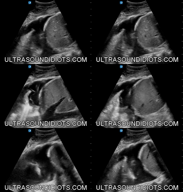
Findings: The is no free fluid identified within the pleural space. There is a large volume of free fluid demonstrated within the subphrenic space, hepatorenal space, and paracolic gutter.
Impression: Positive Exam, Complete Visualization of RUQ
EXAM 24
Probe: Phased Array
Depth: Appropriate
Gain: Appropriate
Findings: Free fluid demonstrated in the pleural space. No free fluid identified within the subphrenic space. The hepatorenal space and paracolic gutter are not completely visualized. There is a benign simple cyst with imperceptible wall (Bosniak 1) in the superior pole of the right kidney.
Impression: Positive Exam, Incomplete Visualization of RUQ
EXAM 25
Probe: Phased Array
Depth: Appropriate
Gain: Too High
Findings: There is no free fluid identified in the pleural space or the subphrenic space. There is trace free fluid demonstrated in the hepatorenal space. There is no free fluid identified in the paracolic gutter.
Impression: Positive Exam, Complete Visualization of RUQ
EXAM 26
Probe: Phased Array
Depth: Appropriate
Gain: Appropriate
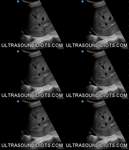
Findings: The exam demonstrates no evidence of free fluid in the pleural space, subphrenic space, hepatorenal space or paracolic gutter. (Great example of normal liver echogenicity: More echogenic than right kidney, good visualization of biliary and vascular system)
Impression: Negative Exam, Complete Visualization of RUQ
EXAM 27
Probe: Phased Array
Depth: Appropriate
Gain: Appropriate
Findings: Free fluid identified in the paracolic gutter. No free fluid identified within the pleural, subphrenic or hepatorenal spaces.
Impression: Positive Exam, Complete Visualization of RUQ
Exams to be read...
You have to be able to read images if you are going to produce images.
Read every ultrasound that radiology reads and review all the images.
