Trauma / EFAsT EXAM Page Editors
Matt Jones, MD, MS, RDMS, RDCS (Emergency Medicine)
DON BYARS, MD, RDMS, RDCS (EMergency Medicine)
A. Kyle Deal, MD (Interventional Radiology)
Jessica Burgess, MD, RDMS (Trauma Surgery)
Image Contributors
Matt Jones, MD, MS, RDMS, RDCS
DON BYARS, MD, RDMS, RDCS
ANNA KATE DEAL, MD, RDMS, RDCS
TAMARA ARMSTRONG, MD
CHARLES GRAFFEO, MD
David Nesbitt, MD
D. Taylor Gammons, MD
Trauma / EFAST Exams
Scroll through the clips and images at your own pace
Reads are posted below images
RUQ EXAM 1-25

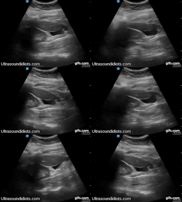
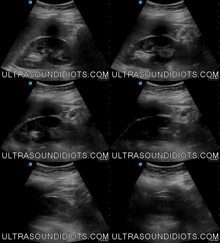
RUQ Exam 1 - Positive Exam, Limited View, Curvilinear probe. Free fluid located below the inferior pole of the right kidney. This location is often overlooked and can be easily missed if the sonographer does not slide the probe inferiorly to visualize the lower pole of the kidney. The the pleural space, the subphrenic space (between diaphragm and liver), and the hepatorenal space (Morrison’s pouch) are not visualized in the clip.
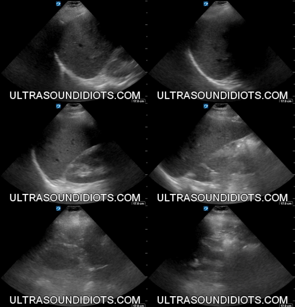
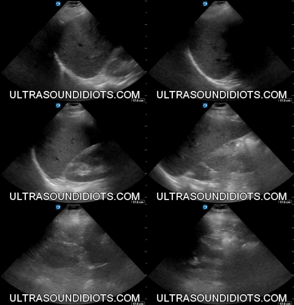
RUQ Exam 2 - Negative Exam. This is an excellent clip which clearly shows all four areas of interest- the pleural space, the subphrenic space (between diaphragm and liver), the hepatorenal space (Morrison’s pouch), and the inferior pole of the kidney.
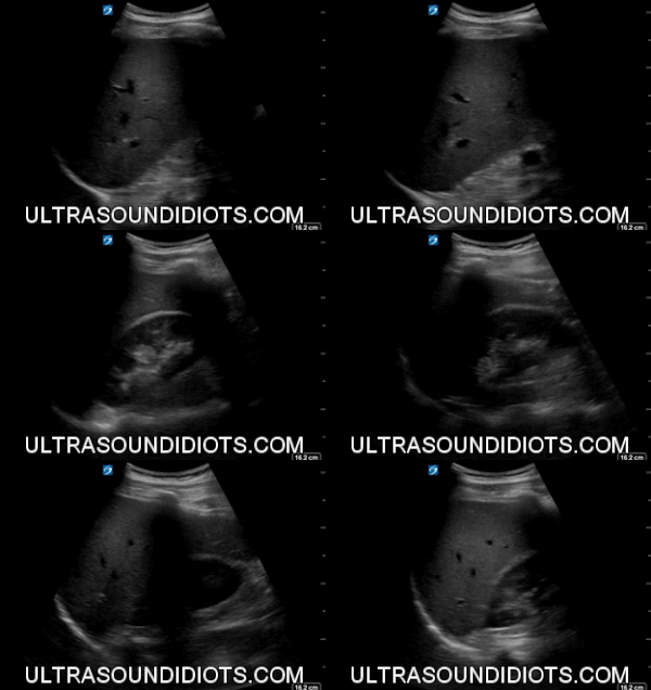

RUQ Exam 3 - Indeterminate Exam. This clip does not adequately visualize the pleural space (space above the diaphragm). Otherwise, the exam shows no evidence of free fluid in the subphrenic space, the hepatorenal space or the inferior pole of the right kidney.
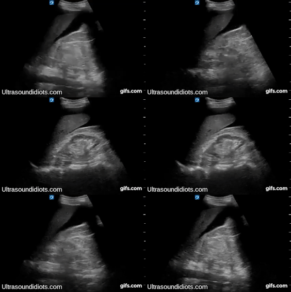

RUQ Exam 4 - Positive Exam, Limited View. Free Fluid located within Morrison's pouch and inferior pole of right kidney. The pleural space and subphrenic space are not visualized.

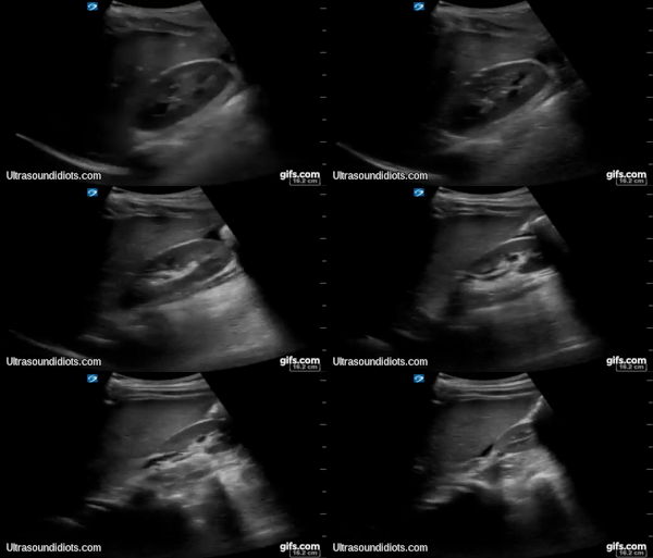
RUQ Exam 5 - Positive Exam, Limited View. Free fluid located within Morrison's pouch. The pleural space, the subphrenic space and the inferior pole of the right kidney are not visualized.

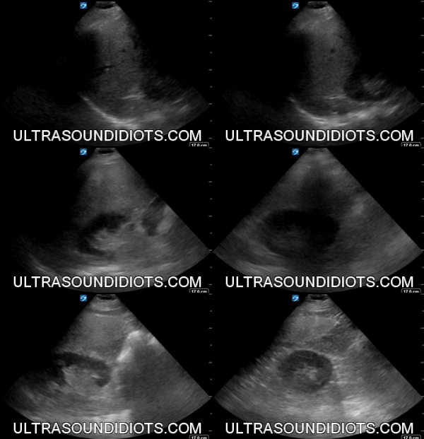
RUQ Exam 6 - Indeterminate Exam. This clip does not adequately visualize the pleural space (space above the diaphragm). Otherwise, the exam shows no evidence of free fluid.

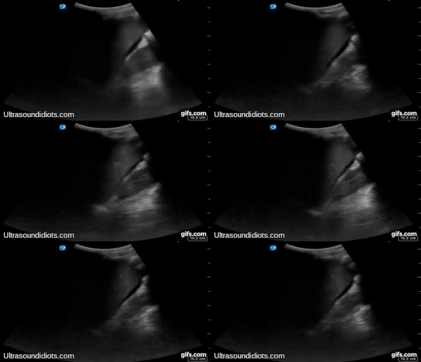
RUQ Exam 7 - Positive Exam, Limited View. Free fluid located within Morrison's pouch. The pleural space, the subphrenic space and the inferior pole of the right kidney are not visualized.

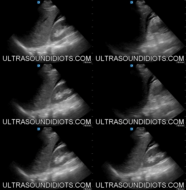
RUQ Exam 8 - Positive Exam, Limited View. Free fluid located within Morrison's pouch. This was a delayed presentation, 2 days after injury. Note the hyperechoic tissue (clot) in Morrison's pouch. The pleural space, the subphrenic space and the inferior pole of the right kidney are not visualized.


RUQ Exam 9 - Positive Exam, Limited View. This exam is tricky. The gallbladder is present at the beginning of the clip and should not be confused with free fluid. Close inspection of the later part of the clip shows free fluid below the inferior pole of the kidney. The the pleural space, the subphrenic space (between diaphragm and liver), and the hepatorenal space (Morrison’s pouch) are not visualized in the clip.


RUQ Exam 10 - Positive Exam, Limited View. Free fluid located within the pleural space consistent with hemothorax in the setting of trauma. The subphrenic space demonstrates no free fluid. Morrison's pouch and the inferior pole of the right kidney are not visualized.
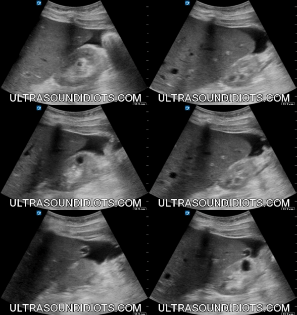

RUQ Exam 11 - Positive Exam, Limited View. Free fluid located below the inferior pole of the right kidney. The the pleural space and the subphrenic space are not visualized in the clip. Note that there is no fluid present in Morrison's pouch.

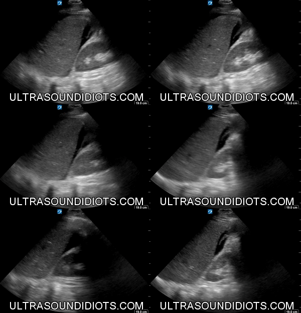
RUQ Exam 12 - Positive Exam, Limited View. Free fluid located within Morrison's pouch. This was a delayed presentation, 2 days after injury. Note the hyperechoic tissue (clot) in Morrison's pouch. The pleural space, the subphrenic space and the inferior pole of the right kidney are not visualized.

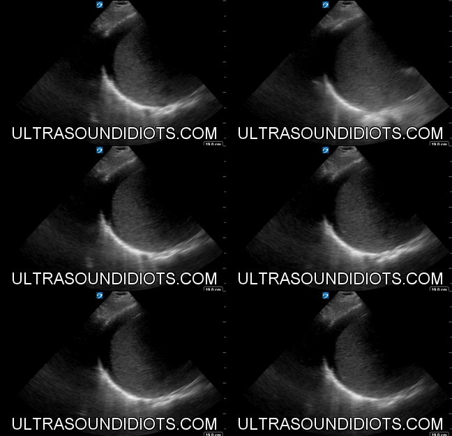
RUQ Exam 13 - Positive Exam, Limited View. Free fluid located in the subphrenic space. There is not fluid in the pleural space. Morrison's pouch and the inferior pole of the right kidney are not viewed.


RUQ Exam 14 - Negative Exam. This is an excellent clip which clearly shows all four areas of interest- the pleural space, the subphrenic space (between diaphragm and liver), the hepatorenal space (Morrison’s pouch), and the inferior pole of the kidney.


RUQ Exam 15 - Positive Exam, Complete View. There is no fluid in the pleural space. Free fluid located in the subphrenic space. The hepatorenal space and the inferior pole of the right kidney demonstrate no free fluid.
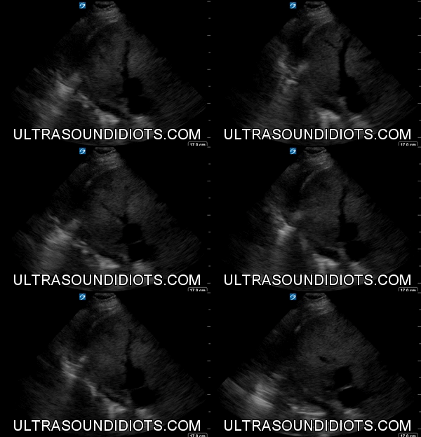

RUQ Exam 16 - Positive Exam, Limited View. There is a small amount of fluid in the pleural space. The subphrenic space demonstrates no free fluid. Morrison’s pouch and the inferior pole of the right kidney are not visualized.

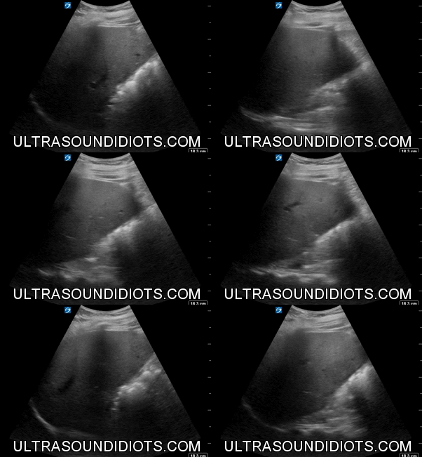
RUQ Exam 17 - Thricky… Indeterminate Exam. The pleural space is not visualized. There is not subphrenic fluid. This patient is s/p rt nephrectomy. There is no fluid visualized in the hepatorenal space. The space that would occupy the inferior pole of the right kidney is not visualized.
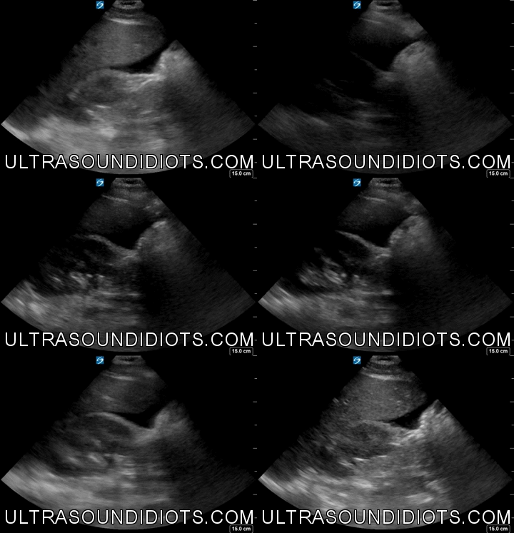

RUQ Exam 18 - Positive Exam, Limited View. The pleural space and the subphrenic space are not visualized. There is fluid in Morrison’s pouch and the inferior pole of the right kidney.


RUQ Exam 19 - Negative Exam. This is an excellent clip which clearly shows all four areas of interest- the pleural space, the subphrenic space, the hepatorenal space, and the inferior pole of the kidney.
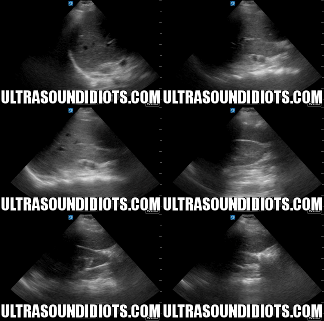

RUQ Exam 20 - Negative Exam. This is an excellent clip which clearly shows all four areas of interest- the pleural space, the subphrenic space, the hepatorenal space, and the inferior pole of the kidney.


RUQ Exam 21 - LUQ Exam 14 - Negative Exam. This is an excellent clip which clearly shows all four areas of interest- the pleural space, the subphrenic space (between diaphragm and spleen), the splenorenal space, and the inferior pole of the left kidney.


Exam 22
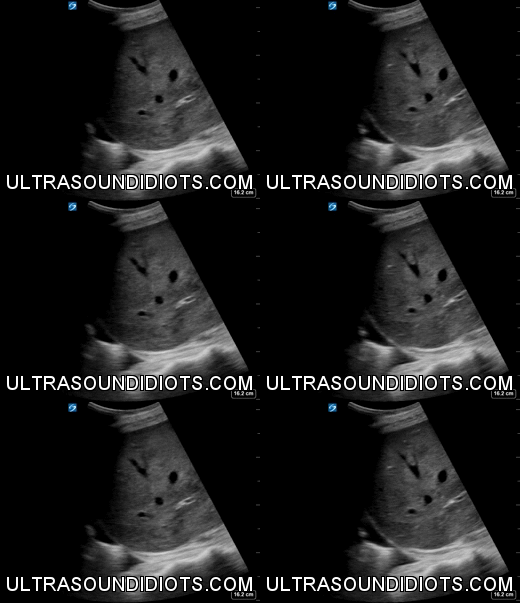

RUQ Exam 23


RUQ Exam 24


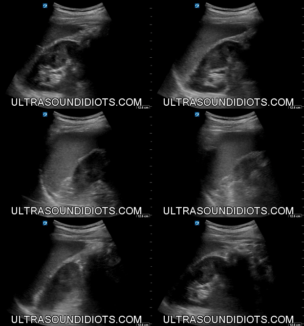

LUQ Exam 1 - Negative Exam. This is an excellent clip which clearly shows all four areas of interest- the pleural space, the subphrenic space (between diaphragm and spleen), the splenorenal space, and the inferior pole of the left kidney.
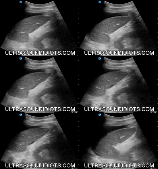

LUQ Exam 2 - Positive Exam, Limited View. The pleural space is not visualized. The subphrenic space has free fluid. There is no fluid in the splenorenal space. The inferior pole of the left kidney is not visualized.
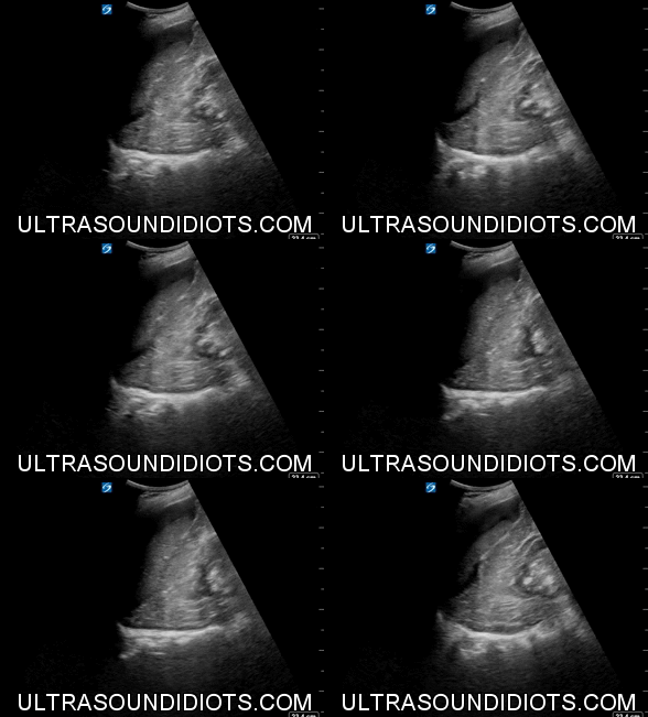

LUQ Exam 3 - Positive Exam, Limited View. The pleural space is not visualized. The subphrenic space has free fluid. There is no fluid in the splenorenal space. The inferior pole of the left kidney is not visualized.


LUQ Exam 4 - Negative Exam. This is an excellent clip which clearly shows all four areas of interest- the pleural space, the subphrenic space (between diaphragm and spleen), the splenorenal space, and the inferior pole of the left kidney.
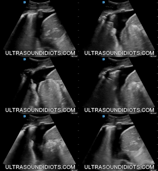

LUQ Exam 5 - Positive Exam, Limited View. Free fluid located within the pleural space consistent with hemothorax in the setting of trauma. The subphrenic space, the splenorenal space and the inferior pole of the left kidney are not visualized.
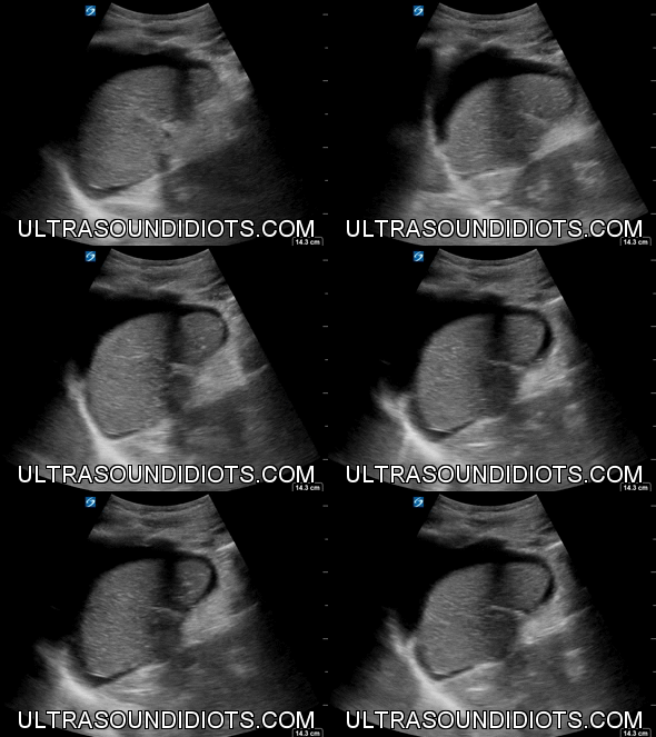

LUQ Exam 6 - Positive Exam, Limited View. The pleural space is not visualized. The subphrenic space has free fluid. The splenorenal space and the inferior pole of the left kidney are not visualized.


LUQ Exam 7 - Negative Exam. This is an excellent clip which clearly shows all four areas of interest- the pleural space, the subphrenic space (between diaphragm and spleen), the splenorenal space, and the inferior pole of the left kidney.
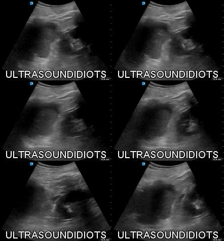

LUQ Exam 8 - Positive Exam, Limited View. The pleural space is not visualized. The subphrenic space has free fluid. There is no fluid in the splenorenal space. The inferior pole of the left kidney is not visualized.
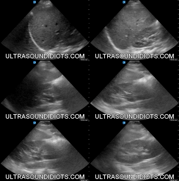

LUQ Exam 9 - Negative Exam. This is an excellent clip which clearly shows all four areas of interest- the pleural space, the subphrenic space (between diaphragm and spleen), the splenorenal space, and the inferior pole of the left kidney.


LUQ Exam 10 - Negative Exam. This is an excellent clip which clearly shows all four areas of interest- the pleural space, the subphrenic space (between diaphragm and spleen), the splenorenal space, and the inferior pole of the left kidney.

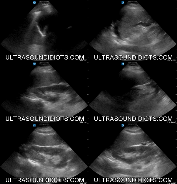
LUQ Exam 11 - Positive Exam, Complete View. There is no fluid in the pleural space. The subphrenic space has free fluid. There is no fluid in the splenorenal space or the inferior pole of the left kidney.
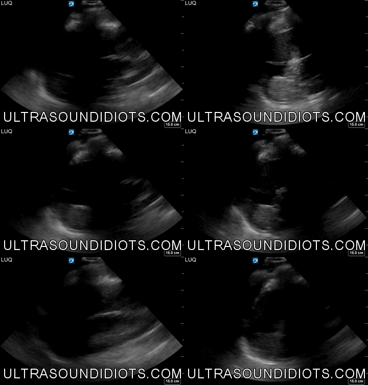

LUQ Exam 12 - Positive Exam, Limited View. The pleural space is not visualized. The subphrenic space is not clear and there does not appear to be free fluid in this space. There is free fluid at the superior pole of the left kidney consistent with fluid in the splenorenal space. The inferior pole of the left kidney is not visualized.
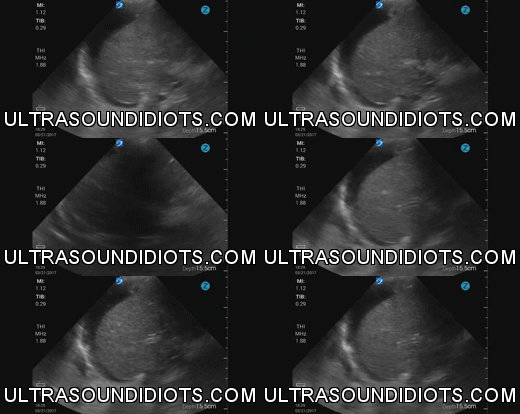

LUQ Exam 13 - Positive Exam, Limited View.There is a small area free fluid above the diaphragm in the pleural space. This is free fluid in the subphrenic space. The splenorenal space and the inferior pole of the left kidney is not visualized.


LUQ Exam 14


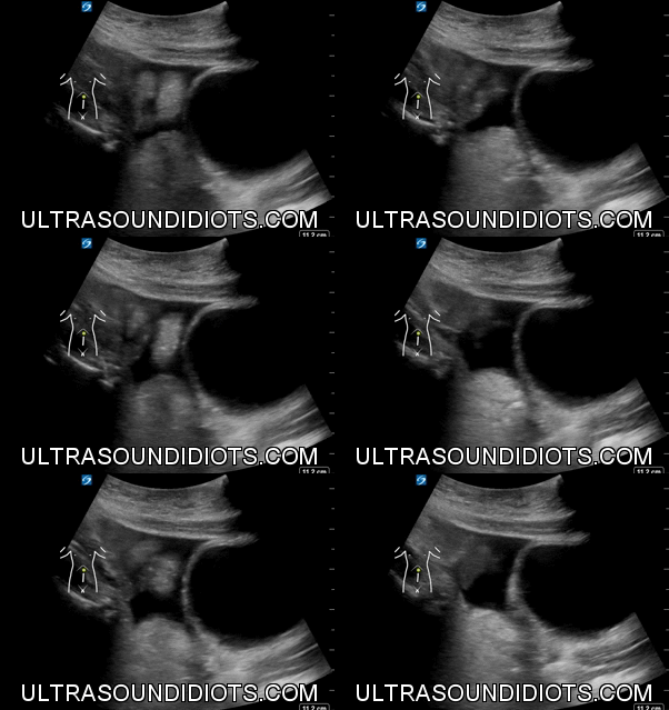
Pelvic Exam 1 - Positive Exam. Sagittal view of the fundus with free fluid above (in the peritoneum).
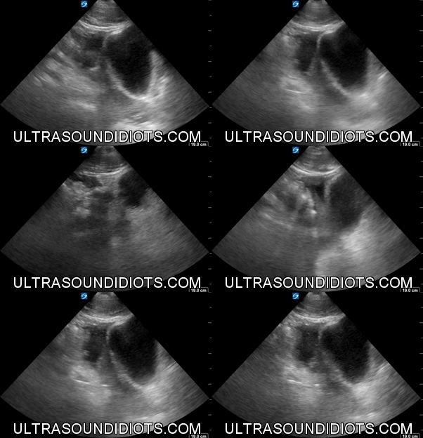
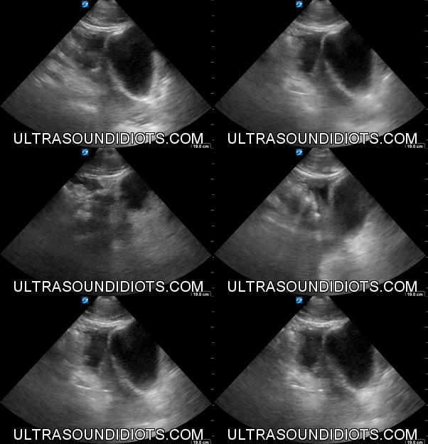
Pelvic Exam 2 - Positive Exam. Sagittal view of the fundus with a small amount of free fluid superior and posterior to the bladder (in the peritoneum).


Pelvic Exam 3 - Positive Exam. Limited View. Transverse view of the fundus with free fluid superiorly (in the peritoneum).

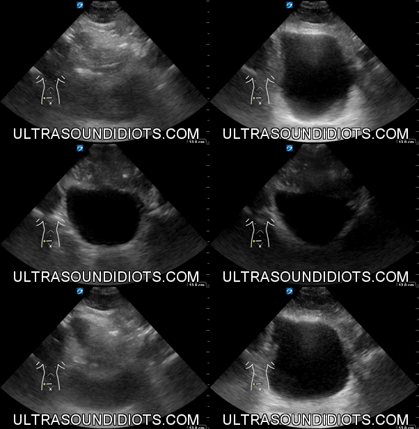
Pelvic Exam 4 - Negative Exam. Transverse view of the bladder fundus with no superior or surrounding free fluid.

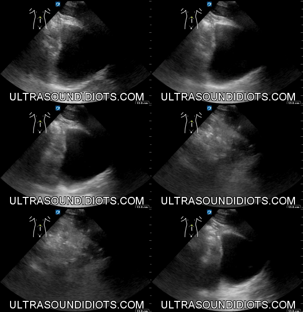
Pelvic Exam 5 - Negative Exam, Limited. Sagittal view of the bladder fundus with no surrounding free fluid seen. This is not a complete study. It is a 3 second clip without clearly showing both sides of the bladder.

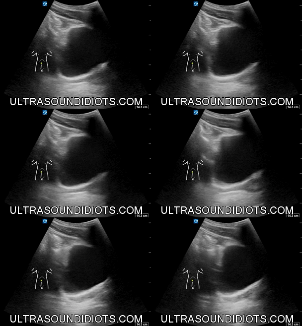
Pelvic Exam 6 - Negative Exam, Limited. Sagittal view of the bladder fundus with no surrounding free fluid seen. This is not a complete study. The sonographer failed to sweep the probe laterally.
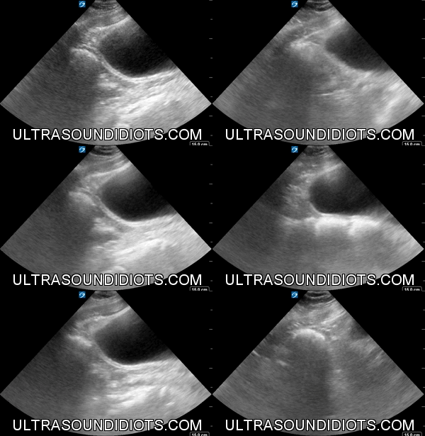

Pelvic Exam 7 - Negative Exam. Sagittal view of the bladder fundus with no surrounding free fluid seen.

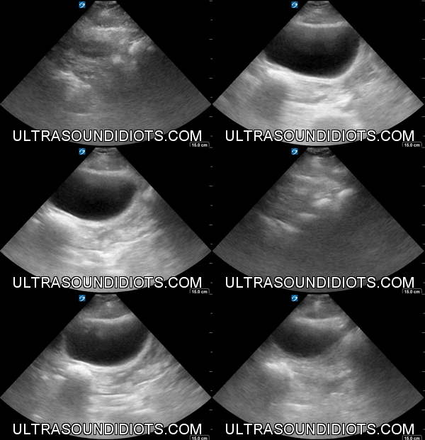
Pelvic Exam 8 - Negative Exam. Transverse view of the bladder fundus with no surrounding free fluid seen.


Pelvic Exam 9 - Positive Exam. The clip starts with transverse view. There is fluid superior to the bladder findus. In the second part of the clip, the sonographer rotates the probe to show a sagittal view of the fundus. There is free fluid superior to the fundus.

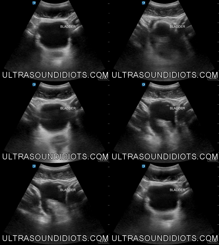
Pelvic Exam 10 - Positive Exam. Transverse view. There is fluid superior to the bladder findus.
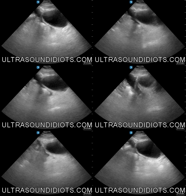


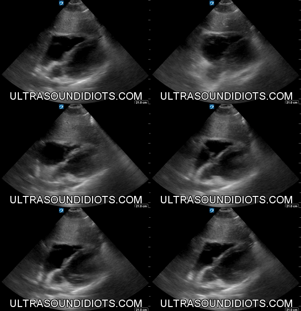
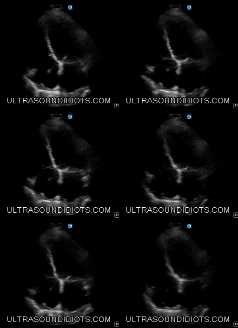
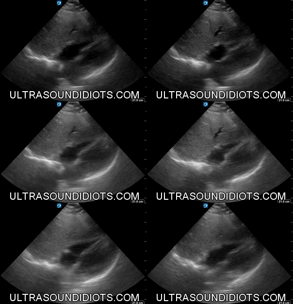
Pericardial Exam 1 - Negative exam. No free fluid visualized within the pericardium.


Pericardial Exam 2 - Negative exam. No free fluid visualized within the pericardium.


Pericardial Exam 3 - Small to moderate pericardial effusion. No evidence of right ventricular dysfunction during diastole.


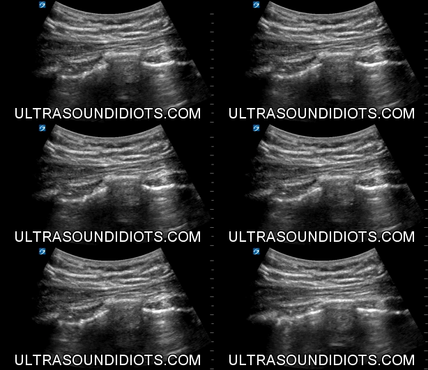

Pleural Exam 1 - Negative for pneumothorax in this view. Unilateral b-mode view of pleural sliding with curvilinear probe.


Pleural Exam 2 - Negative for pneumothorax in this view. Unilateral m-mode view of pleural sliding with curvilinear probe.

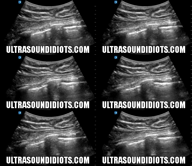
Pleural Exam 3 - Negative for pneumothorax in this view. Unilateral b-mode view of pleural sliding with curvilinear probe.


Pleural Exam 4 - Negative for pneumothorax in this view. Unilateral m-mode view of pleural sliding with curvilinear probe.

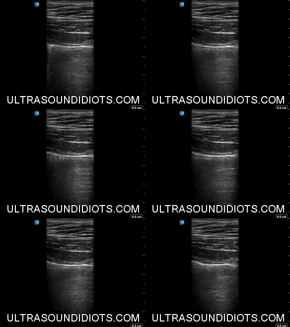
Pleural Exam 5 - Negative for pneumothorax in this view. Unilateral b-mode view of pleural sliding with linear probe.

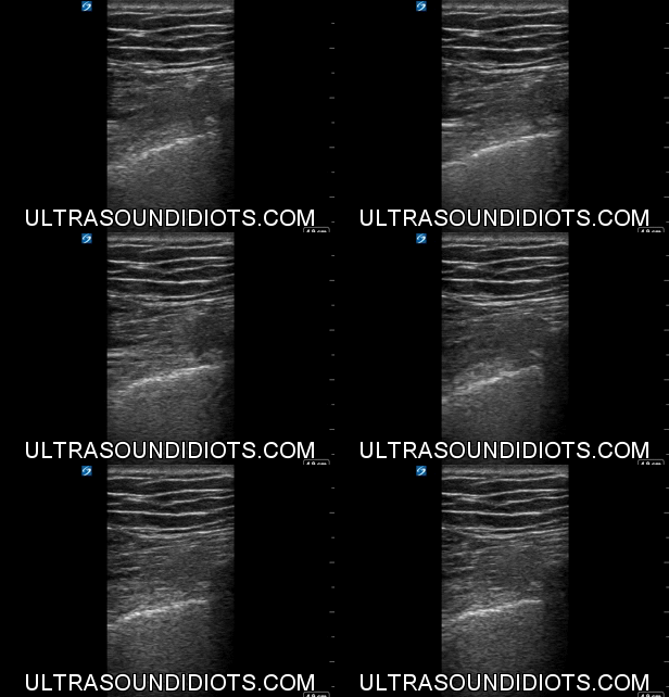
Pleural Exam 6 - Positive Exam. Unilateral B-mode view demonstrating absence of pleural sliding, consistent with pneumothorax.


Pleural Exam 7 - Negative for pneumothorax in this view. Unilateral m-mode view of pleural sliding with linear probe.

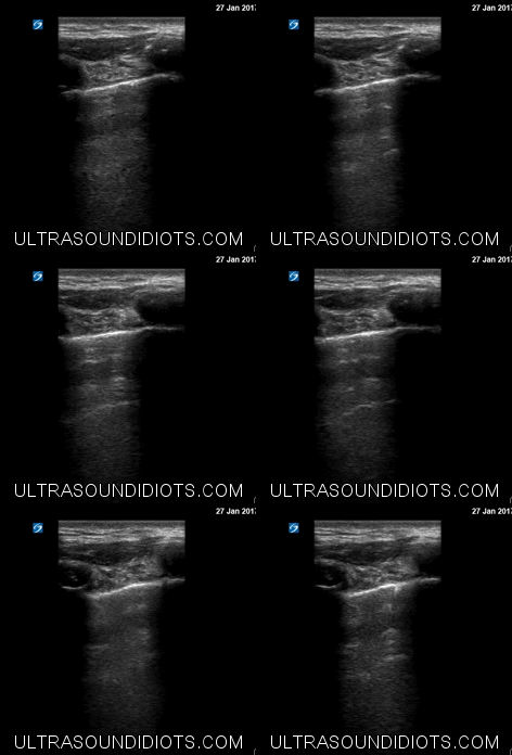
Pleural Exam 8 - Negative for pneumothorax in this view. Unilateral b-mode view of pleural sliding with linear probe.
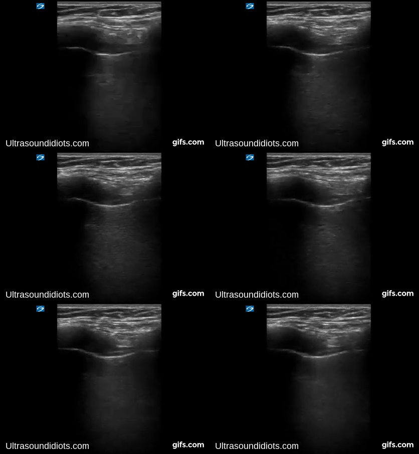

Pleural Exam 9 - Positive Exam. Unilateral B-mode view demonstrating absence of pleural sliding, consistent with pneumothorax.


Pleural Exam 10 - Positive Exam. Unilateral M-mode view demonstrating absence of pleural sliding, consistent with pneumothorax.


Pleural Exam 11 - Negative for pneumothorax in this view. Unilateral b-mode view of pleural sliding with sector probe.


Pleural Exam 12 - Negative for pneumothorax in this view. Unilateral m-mode view of pleural sliding with sector probe.

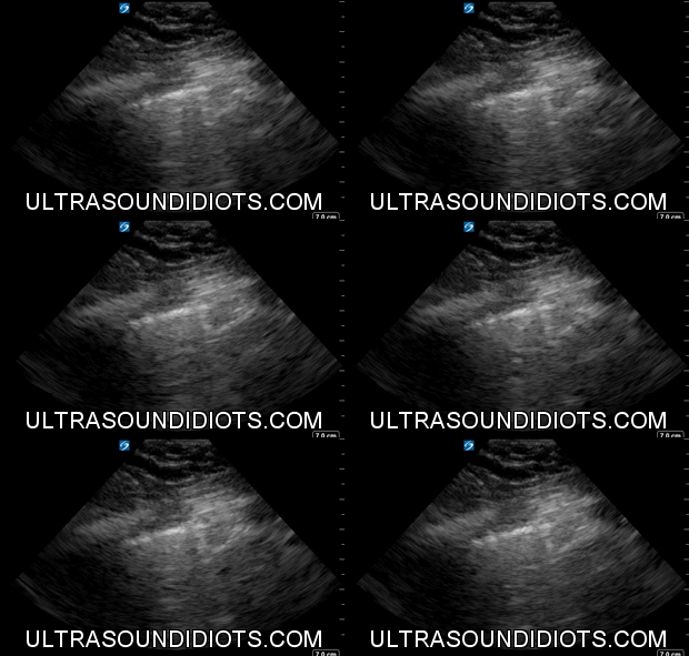
Pleural Exam 13 - Positive Exam. Sector probe. Unilateral B-mode view demonstrating absence of pleural sliding, consistent with pneumothorax.


Pleural Exam 14 - Positive Exam. Sector Probe. Unilateral M-mode view demonstrating absence of pleural sliding, consistent with pneumothorax.

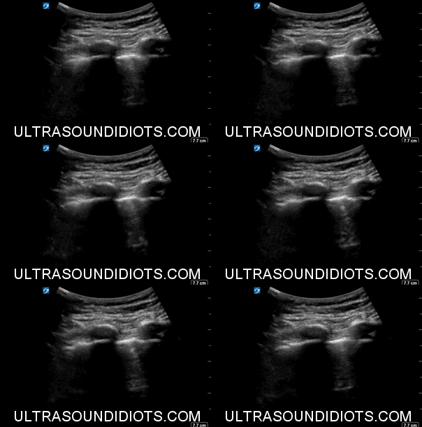
Pleural Exam 15 - Negative for pneumothorax in this view. Unilateral b-mode view of pleural sliding with curvilinear probe.


Pleural Exam 16 - Negative for pneumothorax in this view. Unilateral m-mode view of pleural sliding with curvilinear probe.

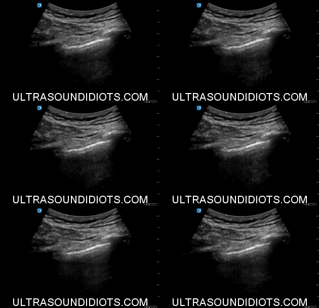
Pleural Exam 17 - Positive Exam. Curvilinear Probe. Unilateral B-mode view demonstrating absence of pleural sliding, consistent with pneumothorax.


Pleural Exam 18 - Positive Exam. Curvilinear Probe. Unilateral m-mode view demonstrating absence of pleural sliding, consistent with pneumothorax.

Pleural Exam 19 - Negative for pneumothorax in this view. Unilateral b-mode view of pleural sliding with linear probe.

Pleural Exam 19 - Negative for pneumothorax in this view. Unilateral b-mode view of pleural sliding with linear probe.


Pleural Exam 20 - Negative for pneumothorax in this view. Unilateral m-mode view of pleural sliding with linear probe.


