Artifacts - Cody
Causes of Artifacts:
Violation of assumptions incorporated into every US system
Sound travels in a straight line.
Sound travels directly to a reflector and back.
Sound travels in soft tissue at 1540m/s.
Reflections arise only from structures positioned in the beam’s main axis
The imaging plane is very thin.
The strength of the reflection is related to the characteristics of the tissue creating the reflection.
Equipment malfunction or poor design
US Physics
Operator Error
Many artifacts will disappear as you change views, whereas anatomy will remain visible regardless
Reverberation
Appear as multiple, equally spaced echoes that occur because the sound bounces between two strong reflectors that are parallel to the US beam.
Each bounce between the reflectors increases the time it takes to return back to the US machine. The machine interprets the increased time to mean that the reflector is deeper. (Assumption 2)
Characteristics:
Appear in multiples and equally spaced
Are parallel to the sound beam's main axis
Located at ever-increasing depths
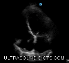
[Example with normal anatomy or pathology]
A lines in the lung -> reverb from pleural line and skin
Comet Tail (ring down artifact)
Reverberations that are closely spaced together almost blending together, usually occur in medium with very high propagation speeds such as prosthetic valve.
Z lines or B lines in the lung are comet tails from reflections between the parietal and visceral pleura which are very close together. B lines are propagated further than Z lines due to the presence fluid allowing deep conduction of the sound waves.
Shadow
Hypoechoic or anechoic region that extends downward from a highly attenuating structure
The structure above is more attenuating than the tissue beneath it
Attenuation occurs via reflections, scatter, or absorption
Assumption 6
Edge Shadow
Sound beam refracts at the edge of a curved reflector. At the same time the beam diverges both of which result in a drop in intensity leading to shadow
Gallbladder or large vessels cause edge shadow [example image]
Assumption 6
Posterior Enhancement
Hyperechoic region beneath a very low attenuating structure such as fluid.
Assumption 6
Cyst, bladder, gallbladder, etc.
Mirror Artifact
When sound reflects off a strong reflector and redirected to a second reflector
The artifact always appears deeper than the true reflector and the bright reflector lies in between the the real image and the artifact
Liver and diaphragm [example image]
Source:
Edelman, Sidney K. Understanding ultrasound physics. 4th ed. Woodlands, TX: ESP, 2012.