Aorta Anatomy
Abdominal Aorta
Two major views include long (sagittal) and short (axial)
Long axis view = dot oriented to head
Short axis view = dot oriented to the patient's right
Curvilinear Probe, low frequency, better for depth
Slowly increasing pressure will displace gas to provide better images
Short Axis View


Long Axis View
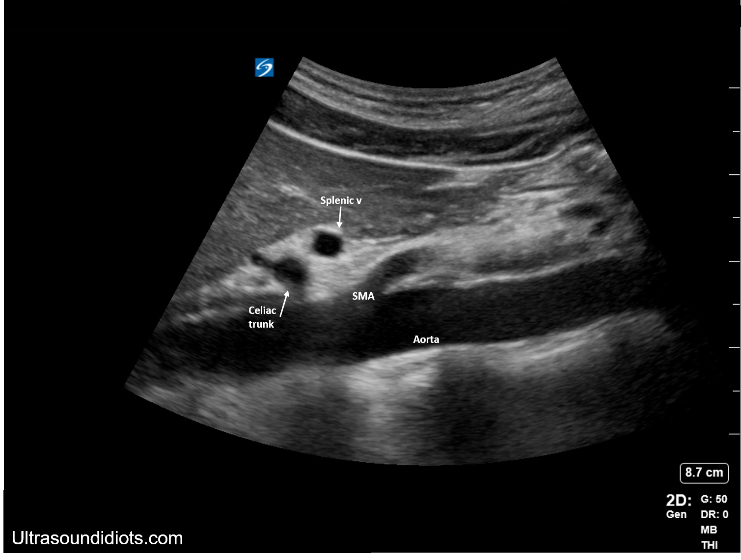
Proximal Abdominal Aorta - demonstrates proximal major vessels of abdominal aorta
Distal Abdominal Aorta - demonstrates distal aorta down to level of bifurcation


Thoracic Aorta
Parasternal long axis view
Right parasternal long axis view
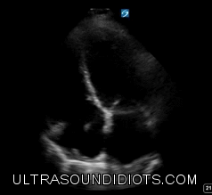
Suprasternal Notch View
EVALUATION OF THE AORTIC ARCH FROM THE SUPRASTERNAL NOTCH VIEW USING FOCUSED CARDIAC ULTRASOUND